


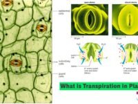
In this tutorial, we have defined ‘What is Fungi?‘ and explored ‘Classification of fungi‘, ‘Economic Importance of fungi’, ‘Affinities‘ etc.
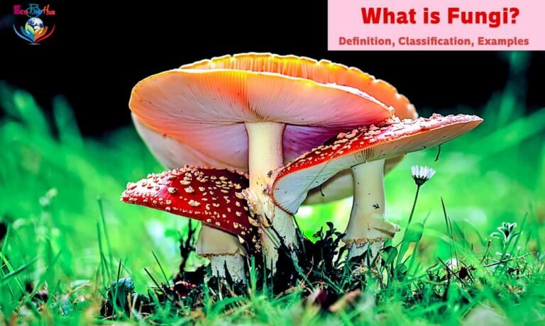
— Fungi Definition: Fungi (singular, fungus) are achlorophyllous, heterotrophic, spore-forming, non-vascular, eucaryotic organisms or plants, which reproduce both sexually and asexually and whose generally filamentous and much branched plant body (mycelium) is surrounded by a cell wall made of chitin or fungal cellulose or both. They possess glycogen as a food reserve. They are cosmopolitan in occurrence being present in the air, water, soil, over and inside animals and plants. They are more abundant in warm and humid areas. Fungi is a large kingdom of over 100,000 species.
— The term fungus is a latin word meaning mushroom.
— Branch of biology dealing with the study of fungi is known as mycology (Gk. mykos = mushroom and logos = discourse) and The branch of biology dealing with fungal diseases and disease causing fungi is known as fungal pathology.
— Clausius (1601) is regarded as the earliest writer who described fungi.
Pier Antonio Micheli (Italian botanist) gave accurate descriptions and illustrations of fungi in his books ‘Nova Planatarum Genera‘ and he is rightly called ‘Father of Mycology‘.
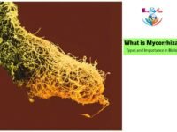
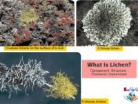
► Read More: Angiosperm Plant Families and their Floral Formula ► Read More: What is Mycorrhizae? Types and Importance in Biology ► Read More: What is Lichen? Component, Structure, Economic Importance ► Read More: Origin of Life (Biopoiesis) and Origin of Universe ► Read More: What is Life? Definition & Characteristics of Life ► Read More: Transportation in Plants || Means of Transport
TABLE OF CONTENTS
CHARACTERISTICS OF FUNGI
— Fungi are ubiquitous i.e occur in a variety of habitats.
— Most of them are moisture loving and terrestrial, but a few are aquatic, e.g., Monoblepharis and Saprolegnia and these are commonly known as ‘water moulds’.
— A few fungi are epiphytic, live on trees, e.g., Armillaria on the apple tree and causes red rot of apple.
— It is usually defined as a group of those organisms that form the thallus (i.e., not differentiated into root, stem and leaves), built up of single cell or cells (unicellular or multicellular).
— Fungi lack chlorophyll and are unable to synthesize their own food by the process of photosynthesis. Fungi obtain their nutrition from the external source by the process of extracellular digestion and absorption of the digested material. Such mode of nutrition is called heterotrophic and the organisms are called heterotrophs.
Heterotrophic organisms either live on dead decaying organic matter or on living organisms.
— According to their mode of nutrition, fungi are of two types – parasites and saprophytes.
● Parasite may be defined as “an organism existing in an intimate association with another living organism from which it derives an essential part of the materials for its existence”. Thus, these organisms grow on living organisms and obtain
their food from it.
Parasitic fungi obtain their food from living hosts. These may be :
– Ectophytic : These are externally on the host, e.g., Erysiphe (Powdery mildew).
– Endophytic : These are inside the tissue of plants, e.g., Albugo, Phytophthora, Alternaria.
● Saprotrophic fungi obtain their organic food requirement from dead and decaying organic matter, fruits, vegetables, meat, etc. Along with certain bacteria, the saprotrophic fungi function as the main decomposers of organic remains. They are essential for the recycling of inorganic resources in the biosphere, i.e., biogeochemical cycling. However, saprotrophs spoil our food. Foods are protected from the attack of saprotrophic bacteria and fungi by keeping them in refrigerators. Because of low temperature in refrigerators bacteria and fungi become largely inactive.
Some fungi live as symbionts in lichens (along with algae) and mycorrhiza (roots of higher plants).
— Fungi possess a definite cell wall (containing cellulose or chitin or both i.e., fungus cellulose) and true nucleus (eukaryotic) but lack chlorophyll (achlorophyllous) and differentiation of vascular tissue (i.e. nonvascular).
— They are spore forming and reproduce by vegetative, asexual and sexual methods.
— The reserve food material is in the form of glycogen and oil globules.
— Fungi grow well at 20-30ºC and at acidic pH (6.0).
— Fungi may be unicellular (yeast), many branches filamentous (Rhizopus) type.
► Fungal Structure and Nature of Growth
Except for yeast, the body filaments known as hyphae (singular, hypha; gk word hypha – web). A mass of connected hyphae is called a mycelium (plural, mycelia). A mycelium is the main body of multicellular fungi that grows into, on, or through soil, wood, or tissues of host organisms. Because of the threadlike nature of hyphae, a mycelium has a high surface-area-to-volume ratio. This means that the absorption of nutrients is very efficient, which also contributes to the high growth rates of some fungi under ideal conditions. When growth conditions are not optimal, reproductive structures can form from dense masses of hyphae. The spores produced by these structures are resistant to adverse conditions, such as desiccation, and germinate when conditions become suitable. These reproductive structures are most obvious in the forms of mushrooms, toadstools, and puffballs.
A mycelium develops from a single spore through branching and apical growth of the first hypha or germ tube.
Yeast does not have a mycelium. Its cells may adhere to form short temporary chains. Such a short temporary chain is known as pseudomycelium.
In yeast, the same cell may function in vegetative growth as well as in sexual reproduction. In most other lungi there are two distinct phases: vegetative and reproductive. The vegetative phase is also called assimilative phase, During this phase there is rapid absorption of nutrients from the substratum, However, the fungus is seldom noticeable due to two reasons : (a) Hyaline nature of hyphae, (h) Occurrence of the major part of mycelium inside the substratum.
Reproductive phase is conspicuous in most of the fungi. The hyphae often become aerial. They form fructifications or fruiting bodies. In some cases the spores produced during this phase are coloured.
► Tissue and Cell Structure
(i) Cell walls contain Chitin
Unlike the cell walls of plants and many protists, fungi have cell walls composed primarily of the polysaccharide chitin. Like the cellulose found in plant cell walls, chitin is a polymer; however, chitin it is made from a modified, nitrogen-containing form of glucose called N-acetylglucosamine. The polymers of N-acetylglucosamine are then cross-linked with proteins. In some fungi cellulose is also present, either alone (e.g., Phytophthora and many other oomycetes) or along with chitin.
Chitin is also found in the exoskeletons of mollusks, insects, and crustaceans The presence of cell walls made of chitin in both animals and fungi is part of the evidence supporting the idea that fungi are more closely related to animals than they are to plants. Additional evidence comes from the observation that both fungi and animals store excess glucose in the form of glycogen; plants, on the other hand, store starch.
(ii) Cell Structure
— Fungal cells have a eucaryotic structure.
— Plastids are, however, absent.
— A membranous vesicle called lomasome is found attached to the plasma membrane.
— Endoplasmic reticulum, vacuoles, mitochondria, ribosomes, microbodies, microtubules, etc. are typical.
— Dictyosomes ( (Golgi bodies) are unicisternal.
— The cytoplasm appear granular and contains many minute nuclei scattered in the peripheral layer.
— Food reserve is in the form of glycogen and oil.
— The vacuoles are small, few or absent in the actively growing tips of the mycelium. Special vesicles having wall materials occur near hyphal tips. They are called chitosomes.
— Nuclei are small and inconspicious as compared to other eukaryotes.
— Cell division is karyochoresis and spindle is intranuclear in both mitosis and meiosis. Spindle poles either contain centrioles or spindle pole bodies (SPB). The latter lack microtubular organization.
(iii) Types of Hyphae
— Hyphae are of two types:
➢ aseptate (= nonseptate) or coenocytic (multinucleate) and
➢ septate (number of partitions or septa).
— Aseptate hyphae may form septa during reproduction, eg Rhizopus, Albugo, Phytophthora etc. In aseptate hyphae, septa are not formed at the time of division. Continued nuclear division makes the hyphae multinucleate. If the whole mycelium is without septa, the same is called coenocytic.
— In septate form cell may be monokaryotic (uninucleate) or dikaryotic (two nuclei) or multinucleate.
— Septa are of 3 types: complete septum, septum with simple pore, and septum with dolipore.
➢ In complete septum the cross wall is complete without distinct pores, e.g., Geotrichum.
➢ In most ascomycetes and deuteromycetes, the septum possesses a simple central pore. A simple central pore may get plugged by membrane bound crystalline structure called woronin body, e.g., trichomycetes. Woronin body is secreted by a microbody and covered by a membrane.
➢ In basidiomycetes, the septum becomes barrel shaped around a central pore called dolipore septum, it may be surrounded by pore cap.
Septal pores allow quick transport of nutrients from the region of absorption to all parts of the mycelium, mobilization of reserve materials from older parts to younger parts, and from vegetative hyphae to reproductive hyphae.
(iv) Hyphae Structure
— In several cases fungal hyphae come together and produce a loose or compact tissue known as plectenchyma (Gk plekein– to weave, enchymatissue), Plectenchyma is of Iwo types: prosenchyma and pseudoparenchyma.
➢ Prosenchyma (Gk. pros– towards, enchyma -tissue) Prosenchyma is the mycelium in which hyphae are loosely interwoven and lie more or less parallel to each other. The cells are elongated and easily distinguishable from one another.
➢ Pseudoparenchyma (Gk, pseudo-false, parenchyma– tissue) is the mycelium in which the hyphae are closely packed. They lose their individuality and cannot be distinguishable from one another. The cells appear to be more or less isodiametric or oval. As such they resemble the cells of plant parenchyma.
— When mycelium by interweaving forms compact resting structure it is called sclerotium.
— When fungal mycelia are interwoven to form thick cordlike structures like roots, that help in absorption, it is called rhizomorphs.
— When the plant body is unicelled at one stage and mycelial at the other end then the organisation is described as dimorphic.
► Some cells (or hyphae) contain Multiple Nuclei
Fungi are different from most animals and plants in that each cell (or hypha) can house one, two, or more nuclei. A hypha that has only one nucleus is monokaryotic; a cell with two nuclei is dikaryotic. In a dikaryotic cell, the two haploid nuclei exist independently. Dikaryotic hyphae have some of the genetic properties of diploids, because both genomes may be transcribed.
Sometimes many nuclei are found in the common cytoplasm of a fungal mycelium, which can lack distinct cells. If a dikaryotic or multinucleate hypha has nuclei that are derived from two genetically distinct individuals, the hypha is called heterokaryotic. Hyphae whose nuclei are genetically similar to one another are called homokaryotic.
► Reproduction
— When the entire mycelium is converted into reproductive structure, the thallus is called holocarpic.
— When a part of thallus become reproductive, the thallus is called eucarpic. This can be monocentric (have single sporangium) or polycentric (have many sporangia).
Fungi reproduce by three methods— vegetative, asexual and sexual.
(i) Vegetative Reproduction
It occurs by fragmentation, budding, fission, sclerotia, and rhizomorphs.
01. Fragmentation: The mycelium breaks up into two or more segments due to mechanical injury, decay or some other reason. Each fragment grows to form the complete mycelium.
02. Budding: Some fungi produce small outgrowths, i.e., buds from their vegetative body and receive one daughter nucleus from parent nucleus through division. After sometime, the buds are cut off and mature to form new individuals e.g., yeast and Ustilago.
03. Fission: Splitting of the cell into two daughter cells by constriction, e.g., yeast.
04. Sclerotia: They are perennating bodies made up of compact masses of hyphae. They grow under favourable conditions to produce new mycelia singly, e.g., Claviceps or may be produced in large number from one mycelium in Botrytis.
05. Rhizomorphs: They are rope-like twisted subterranean masses of hyphae with well defined apical growing point. They pass the unfavourable periods in dormant stage. Under favourable conditions each rhizomorph gives rise to a new mycelium.
(ii) Asexual Reproduction
It occurs through the formation of spores. Spores are single celled propagules
Meiospores are more important because prior to their formation, there has been a chance combination of genes during fertilization and chance segregation of genes during meiosis and therefore, meiospores show genetic variations. On the other hand, mitospores are true to the parental characters. They do not add to the variability.
01. Zoospores: They are motile spores that occur in some phycomycetes, e.g., Phytophthora, Albugo, Achlya. The spores are commonly naked. 1–2 flagella (uniflagellate or biflagellate) are borne anteriorly, posteriorly or laterally. Flagella help the zoospores to swim in aquatic habitat for proper dispersal.
Biflagellate zoospores are of
➢ Pear-shaped or pyriform, with 2 flagella placed at anterior end, are known as primary zoospores.
➢ Kidney-shaped or beanshaped, bearing two oppositely directed flagella inserted laterally in a furrow or concave side (secondary zoospores).
02. Sporangiospores: They are nonflagellate spores that develop inside sporangia, e.g. Mucor, Rhizopus. Sporangiospores are usually dispersed by air currents. Therefore, they are produced in large numbers.
03. Chlamydospores: They are thick-walled perennating spores which develop at place, along the hyphae by accumulation of protoplasm, rounding off and secretion of thick wall.
04. Oidia: They are formed under conditions of excess water, sugar and certain salts. Oidia are individual cells separated from hyphae. They multiply by budding. Oidia often take part in fermentation, e.g., Rhizopus.
05. Conidia: They are nonmotile exogenous spores which develop through abstriction on the tips or sides of special hyphae called conidiophores, e.g., Pencillium, Aspergillus,
06. Ascospores: They are a type of nonmotile meiospores which are produced inside special sacs called asci (singular- ascus). An ascus often contains 8 ascospores because meiosis is accompanied by one mitosis. Ascospore formation is characteristic of class ascomycetes.
07. Basidiospores: Basidiospores are nonmotile meiospores which are formed exogenously on short outgrowths of club-shaped structure called basidium. Basidiospores are characteristic of basidiomycetes.
08. Binucleate Spores: The spores are meant for multiplying the dikaryotic mycelium. They are, therefore, themselves dikaryotic, e.g., aecidiospores, uredospores. Both these types of spores are found in rusts, e.g., Puccinia or Wheat Rust. Another type of dikaryotic spore is teleutospore or teliospore. It is specialized to produce a basidium.
Zoospores are useful tools of dispersal in aquatic habitats. Most fungal spores are nonmotile and small. They are dispersed by wind. Usually spores are formed in large number so that at least a few of them reach favourable substrata. This can be proved by laboratory culture of Rhizopus. A slice of bread is moistened and placed on a moist brick kept in a dish having water. Bread slice is exposed to air for some time. It is then covered by a bell jar to prevent drying up. Within 4–5 days the bread slice will be found to have fine cottony masses which develop blackish spots in the next 2–3 days.
(iii) Sexual Reproduction
— In involves the formation and union of two gametes or their nuclei. Sexual reproduction is absent in the artificial group of fungi called fungi imperfecti or deuteromycetes.
— Depending upon the compatibility in sexual reproduction, fungi are of two types— homothallic and heterothallic.
➢ In heterothallic forms sexual reproduction involves fusion between two genetically different mating types.
➢ In homothallic forms fusion occurs between genetically similar types.
Fusion involves the union of cytoplasms as well as nuclei. The fusion of cytoplasm is called plasmogamy while the fusion of two haploid nuclei is called karyogamy. In higher fungi, karyogamy is delayed and occurs just before meiosis.
In the stage intervening between plasmogamy and karyogamy the cells often contain two nuclei or dikaryons (n + n). Such cells are called dikaryotic cells. The phase is known as dikaryophase. In such fungi, the life cycle is completed in three phases instead of two— haplophase, dikaryophase and diplophase (2n). Meiosis occurs in diplophase.
Meiosis, a reduction division takes place in the zygote, reducing the number of chromosomes to half. The two fusing protoplasts are called sex cells or the gametes. The sex cells are produced within the cells of the thallus called gametangia.
➢ If the fusing gametes are morphologically indistinguishable, then they are called isogametes, and the process of fertilization is called isogamous.
➢ If the fusing gametes differ in size and structure, they are called heterogametes and the process is called heterogamous. Heterogamous type of sexual reproduction is of two types: anisogamous and oogamous.
● Anisogamy consists of the fusion of two morphologically similar gametes which differ in size.
● In oogamy, the gametes are produced inside the morphologically differentiated gametangia. The male gametangium is smaller in size and is called the antheridium, while the female gametangium is larger and is called the oogonium or ascogonium.
— Fungi show progressive reduction in sexuality. Sexual reproduction occurs by five methods:
01. Planogametic Copulation: Flagellate gametes differentiate and fuse. Fusion may be isogamous or heterogamous. Heterogamous fusion may be by anisogamy (gametes different in size and structure) or oogamy. In oogamy, the male gamete is generally small and flagellate while the female gamete is large, food laden and nonflagellate.
02. Gametangial Contact: There are two types of sex organs, male antheridia and female oogonia. Male gametes or nuclei are transferred to the oogonium through a fertilization tube. E.g., Phytophthora, Albugo.
03. Gametangial Copulation: It is a type of conjugation in which both the types of gametangia fuse directly resulting in the formation of a zygospore. E.g., Mucor, Rhizopus.
04. Spermatogamy: In spermatization, some fungi produce numerous, minute, uninucleate, spore-like bodies called spermatia. These are transferred through various agencies to receptive hyphae or trichogyne of female gametangium. Finally, the contents of spermatium are transferred into receptive hypha through a pore. E.g., Puccinia graminis.
05. Somatogamy: Distinct sex organs are absent. Sexual reproduction involves the fusion of two hyphae or cells. E.g., Agaricus.
Homothallism is the condition whereby thalli are morphologically and physiologically identical, so that fusion can occur between gametes produced on the same thallus.
Heterothallism is the phenomenon in which the fusing gametes belong to two genetically distinct strains of the same species though there may not be any morphological distinction between the gametes or structures bearing them. The two sexual strains or mating types are termed as (+), and (-). It was first discovered by Blakeslee in 1904.
CLASSIFICATION OF FUNGI
A number of criteria are used for classifying fungi. The important ones are (a) Morphology or form structure and appearance of fungus. Morphology of assimilative (vegetative) mycelium is useful in only a few cases. Morphology of reproductive structures exhibits more variations and is hence important in fungus classification. (b) Types of spores and their dispersal. (c) Life cycle. (d) Physiology. (e) Biochemistry. The major groups of fungi are following:
I. PHYCOMYCOTA OR PHYCOMYCETES – Lower or Algal Fungi
— Fungi are characterized by aseptate coenocytic hyphae. They are algal fungi and are aquatic fungi and are known as water mould.
— Two types of flagella are present in Phycomycetes: whiplash and tinsel type.
— The life cycle of a phycomycetous fungus consists of an asexual phase and sexual phase.
— Asexual reproduction occurs by spores such as zoospores (motile spores) or aplanospores (non-motile spores) produced endogenously inside sporangia.
— Sexual reproduction is isogamous or heterogamous. Heterogamous reproduction is of two types, anisogamous and oogamous.
— Phycomycota or Phycomycetes is divisible into two groups. oomycetes (e.g., Phytophthora, Albugo) and zygomycetes (e.g., Rhizopus, Mucor).
(i) OOMYCETES — The Oogamous Fungi
01. The mycelium is coenocytic (multinucleate and aseptate).
02. Hyphal wall contains cellulose and other glucans in many members. In some case chitin or fungus cellulose is also present.
03. Asexual reproduction involves the formation of spore containing sacs or sporangia. In aquatic conditions the sporangia produce zoospores. In terrestrial conditions the sporangia often behave as spores, equivalent to conidia. Because of it, the sporangia are often called conidiosporangia.
04. Zoospores are generally biflagellate with heterokont flagellation in which one flagellum is smooth while the other is of tinsel type (having fine surface outgrowths called mastigonemes).
05. Gametes are usually nonflagellate.
06. Sexual reproduction is by gametangial contact in which the male sex organs or antheridium pass its product into the female sex organ or oogonium through a fertilization tube.
07. The product of sexual reproduction is oospore.
■ Examples
01. Late Blight: Phytophthora infestans causes late blight of Potato and occasionally of Tomato as well. Blight is the appearance of brownish to black dead areas. They are first formed on the margins and tips of leaflets. Later on, the whole foliage becomes blighted. Tuber yield is reduced. The surface of the tubers also shows blighting. Irish famine of 1845–1847 was caused by the late blight of Potato
02. White Rust: It occurs in crucifers and is characterized by the appearance of irregular white blisters containing conidiosporangia on the leaves and stems. White rust is caused by Albugo candida (= Cystopus candidus).
03. Damping off: Pythium debaryanum kills seedlings of a number of plants through the collapse of the stem just above the ground level.
04. Downy Mildew: The pathogen produces a cottony or woolly bloom on the surface of the host. Sclerospora graminicola spreads downy mildew in cereals and green ear disease of Pennisetum typhoides (vern. Bajra). Peronospora parasitica causes downy mildew in a number of plants, e.g., Pea, Mustard, Spinach, Onion, etc.
(ii) ZYGOMYCETES — The Conjugation Fungi
01. It is a class of terrestrial fungi that are mostly saprotrophic, rarely parasitic.
02. The mycelium is coenocytic (multinucleate, aseptate).
03. Hyphal wall contains chitin or fungus cellulose.
04. Motile cells (zoospores and planogametes) are absent.
05. Mitospores are nonmotile. They are called sporangiospores as the spores are formed inside sporangia borne at the tips of special hyphae called sporangiophores.
06. Sexual reproduction occurs through gametangial copulation or conjugation. Because of it, zygomycetes are also called conjugation fungi.
07. The gametes are multinucleate and are called coenogametes.
08. Sexual reproduction produces a resting diploid spore called zygospore. Because of the presence of zygospore, the group of fungi is called zygomycetes. Zygospore differs from oospore in that during its formation a distinct large food laden nonmotile female gamete is not produced.
09. Zygospore does not give rise to new mycelium directly. Instead, it produces a new sporangium called germ sporangium (previously called zygosporangium). Germ sporangium forms meiospores called germ spores.
■ Examples
01. Squirting Fungus: Pilobolus crystallinus is a coprophilous or dung mould in which mature sporangia are thrown away upto a distance of 2m.
02. Rhizopus and Mucor: Rhizopus stolonifer (= R. nigricans) is popularly known as black bread mould. Mucor caninus or M. mucedo is coprophilous. It is also called dung mould. Rhizopus and Mucor are the common saprotrophic fungi that attack a variety of food stuffs. Soft rot or leak disease of Strawberry, Apple, Sweet Potato, etc. is due to Rhizopus. Mucor pusillus causes infection of internal organs in human beings. Absidia corymbifera causes bronchomycosis.
Both Rhizopus and Mucor species (e.g., Rhizopus oryzae, Mucor javanicus) are used in alcoholic fermentation. The two also produce a number of organic acids like citric acid, lactic acid and fumaric acid.
➢ Rhizopus stolonifer is one of the most common representative species of Rhizopus. It lives as a saprophyte and is frequently found producing white cobwebby mycelium over the surface of moist bread and other foodstuffs. ➢ The vegetative body is a coenocytic mycelium which forms white and cottony growth on the substratum. ➢ The mycelium is differentiated into three kinds of hyphae – rhizoids, stolons, sporangiophores. ➢ The rhizoids secrete digestive enzymes which convert complex carbohydrates (starch) of the substratum into simple sugars. These sugars are absorbed by the rhizoids. ➢ During favourable conditions, asexual reproduction takes place by the formation of aplanospores. ➢ The erect hyphae for asexual reproduction are known as sporangiophores. ➢ The dome shaped sterile region is known as columella. The peripheral region containing many nuclei is known as sporoplasm. ➢ The spores are non-motile (aflagellate) and hence are known as aplanospores or sporangiophores. ➢ Sexual reproduction occurs by the conjugation of two gametangia. In heterothallic species, e.g., R. stolonifer, sexual reproduction occurs when two hyphae of opposite strains come in close proximity to each other. ➢ The wall between the two gametangia dissolves, their protoplasmic contents mingle and the opposite nuclei fuse in pairs to form a multinucleate zygospore. The zygospore germinates on the return of favourable conditions. Life History of Rhizopus Rhizopus stolonifer (= R. nigricans) is the common black bread mould which is found growing on stale bread in warm and moist seasons. It is saprotrophic (= saprobic = saprophytic) and grows on a variety of organic foods rich in carbohydrates. The body or mycelium of the fungus has four types of nonseptate filaments or hyphae — rhizoidal, stoloniferous, sporangiophores and zygophores. All of them develop from common points called holdfasts or apparent nodes. The rhizoidal hyphae are branched. They penetrate the substratum, excrete digestive enzymes and absorb the digested food. The stoloniferous hyphae are subaerial, arched and unbranched. They spread the mycelium over the substratum. Sporangiophores are unbranched aerial hyphae which appear during asexual reproduction. Zygophores are subaerial hyphae that take part in sexual reproduction. The hyphae are nonseptate and multinucleate. Nuclei are small and numerous. Septa are produced only in connection with the formation of reproductive organs and to separate areas of injury and senescence. The nonseptate and multinucleate mycelium is called coenocyte. ►Vegetative reproduction occurs through fragmentation. ► Asexual reproduction is carried out by three types of mitospores— sporangiospores, chlamydospores, and oidia. 01. Sporangiospores: They are formed under favourable conditions. 2–5 sporangiophores or aerial hyphae develop from a holdfast opposite the rhizoidal hyphae. The tip of each sporangiophore develops a swollen sac called sporangium. Internally sporangium is separated from the rest of the sporangiophore by a dome-shaped partition called columella. A sporangium produces hundreds of non-motile haploid spores called sporangiospores. The sporangiospores are dispersed by air currents. The sporangiospores of Rhizopus are minute, brownish-black and multinucleate. After falling on a suitable substratum each sporangiospore gives rise to a new mycelium. 02. Chlamydospores: They are perennating thick walled spores which are formed on the approach of dry conditions. Thin walled chlamydospores, called gemmae, are produced under less desiccating conditions. On the return of favourable conditions, each chlamydospore gives rise to a new mycelium. 03. Oidia: In a liquid medium rich in sugar and acidic pH, the hyphae become septate and form oidia. Oidia multiply by budding. Oidia take part in fermentation, forming alcohol and organic acids. When transferred to a solid substratum, each oidium forms a new mycelium. ► Sexual Reproduction: Rhizopus stolonifer is heterothallic with two mating types, (+) and (-). When the mycelia of both the mating types are present in the same medium, they produce trisporic acids. Trisporic acids stimulate the mycelia to form a special type of subaerial hyphae called zygophores. Zygophores of (+) and (-) strains come in contact at a number of places. Club shaped progametangia are produced by both the zygophores in the regions of contact. The terminal part of each progametangium separates as a gametangium from the rest called suspensor. The protoplast of each gametangium functions as a multinucleate gamete coenogamete. The common wall between the two gametangia dissolves and the two coenogametes fuse forming first a diploid zygote and then a resting spore called zygospore. Zygospore is dark brown or black rounded spore with a warty surface. Its protoplast contains reserve food and one (according to some) to several diploid nuclei. The latter undergo meiosis at the time of zygospore germination. This produces haploid nuclei of both the mating types but only one nucleus survives. The rest degenerate. The surviving haploid nucleus divides mitotically a number of times to form numerous haploid nuclei of only one strain (+ or – but not both). The protoplast now swells up. The zygospore wall breaks. The swollen protoplast comes out as a hypha called promycelium. A sporangium develops at the tip of the promycelium. It is termed as germ sporangium. Formerly, it was called zygosporangium. The germ sporangium develops uninucleate haploid spores or meiospores named germ spores. The latter are dispersed by air currents. On germination, each germ spore gives rise to a new mycelium. 01. Ascomycetes (Gk. askos- sac, mykes- fungus) is a class of diverse fungi numbering over 30,000 species (Ingold, 1967). They are commonly called sac fungi. They include pigmented moulds (brown, green, blue, pink), powdery mildews, yeasts, cup fungi, morels and truffles. Nutritionally they are saprotrophic, decomposers, coprophilous or parasitic. 02. Ascomycetes are characterised by well developed thallus and production of ascospores. 03. Cell in ascomycetes are uninucleate or multinucleate. 04. The mycelium consists of septate hyphae. Yeasts are an exception in that they are basically unicellular. They may, however, form short temporary filamentous structure called pseudomycelium. 05. The septa possess central pores called septal pores. The pores allow communication between adjacent cells. Septal pores show plugging of different types. 06. Cell wall contains chitin or fungus cellulose. 07. Motile structures do not occur in the life cycle. 08. In yeasts, asexual reproduction occurs through budding and fission. Oidia stage, similar to yeast, is found in some other ascomycetes as well. 09. In the majority of ascomycetes, the common mode of asexual reproduction is through the formation of conidia (singular, conidium). Conidia are nonmotile fungal mitospores which are produced exogenously from the tips and sides of hyphae called conidiophores. Conidia are often coloured brown, green, blue or pink. They provide colouration to the fungus. Greenish and bluish growth on bread, citrus fruits and old leather is due to moulds belonging to ascomycetes e.g., Penicillium, Aspergillus. 10. Conidiophores may be branched or unbranched, scattered or aggregated to form structures like acervulus, synnema, sporodochium, etc. 11. Sexual reproduction takes place through fusion of sex cells, somatic cells, gametangial contact between an antheridium and ascogonium, and autogamy. 12. Fertilization occurs in two steps: plasmogamy and karyogamy. Karyogamy is delayed after plasmogamy. A new transitional phase appears in the life cycle. It is called dikaryophase. The cells of dikaryophase are called dikaryotic cells. Each such cell possesses two nuclei (n+n). 13. Some dikaryotic cells function as ascus mother cells. The latter act as the seats of both karyogamy and meiosis. This converts the cells into asci (singular – ascus). 14. Ascus is a sporangial sac peculiar to ascomycetes. 4–8 haploid meiospores named ascospores are produced internally in each ascus. In most cases, half the number of ascospores belong to one mating type while the other half belongs to the second mating type. 15. The asci may occur freely or get aggregated with dikaryotic mycelium to form fructifications called ascocarps. There are 4 types of ascocarps : ● Cleistothecium: A more or less globose ascocarp which has no natural opening, e.g., Penicillium. ● Perithecium: A globose or flask-shaped ascocarp with an apical opening or ostiole, e.g., Claviceps. ● Apothecium: A cup or saucer shaped ascocarp, e.g., Peziza. ● Ascostroma: In some ascomycetes, the asci are Deuteromycetes formed in cavities, called locules in a stromatic fruiting body, known as an ascostroma, e.g., Pleospora. ■ Examples 01. Yeasts They are frequently found growing saprophytically on substrates that contain sugar, such as decaying vegetables, ripe fruits and grains, sugary exudates of trees and nectar of flowers.
II. ASCOMYCETES — The Sac Fungi

Yeasts are a group of nonmycelial or pseudomycelial ascomycetes which multiply asexually by budding or fission and where asci are not organised into ascocarps. Depending upon the mode of asexual reproduction, yeasts are of three types— budding yeasts (e.g., Saccharomyces), fission yeasts (e.g., Schizosaccharomyces) and halobial yeasts (both budding and fission, e.g., Saccharomycoides). Yeasts in which ascus formation is known are named as true yeasts. Related forms which resemble yeasts in most characteristics but where ascus formation is not reported, are called false yeasts, e.g., Candida, Mycoderma, Cryptococcus. They are otherwise included amongst deuteromycetes.
Yeasts are basically unicellular. Under conditions of rapid growth, they form temporary chains or pseudomycelia. The cells are spherical to cylindrical in outline. The cell wall contains chitin and mannan β-glucan. Nucleus lies at one end of a large central vacuole. Fine dark staining threads of unknown significance extend from nucleus to surface of the vacuole. Nutrition is saprotrophic, rarely parasitic. Yeasts are facultative aerobes, i.e., anaerobes capable of respiring aerobically as well. Asexual reproduction is by budding, fission or both.
Embedded in the cytoplasm, are the mitochondria, endoplasmic reticulum and ribosomes. The reserve food materials in the cytoplasm are in the form of glycogen, oil globules and protein particles.
As it lacks chlorophyll it is saprophytic on the substrate, it secretes enzymes & these enzymes are collectively known as zymase. It helps in fermentation.
Sexual reproduction occurs through conjugation between two cells or two ascospores. Distinct mating types (+ and -) are known in Saccharomyces cerevisiae (Baker’s yeast). Conjugation does not produce dikaryotic cells except occasionally in Saccharomyces cerevisiae. The diploid cells may multiply but ultimately they function as asci producing 4–8 ascospores. Ascospores grow to form haploid vegetative cells or fuse to produce diploid cells.
The three types of life cycle are noted in yeast :
● Haplobiontic: In this type of life cycle, the diploid phase is very short and is confined only to zygote which undergoes meiosis immediately after karyogamy, while haploid phase is very much elaborated e.g., Schizosaccharomyces.
● Diplobiontic: In this type of life cycle which is represented by Saccharomyces ludwigii, the diploid phase is long while haploid phase is very short. Here four ascospores are developed.
● Haplo-diplobiontic: In this type of life-cycle, the haplo and diplo phases are equally well represented showing distinct alternation of generations. This type of life-cycle is represented by Saccharomyces cerevisiae.
► Economic Importance
(i) Brewing Industry: Under anaerobic conditions, sugary solutions inoculated with yeasts are converted into alcoholic beverages, e.g., beer, wine, cider, toddy. They are concentrated further to produce rum and whisky. The two common yeasts used by brewing industry are Saccharomyces cerevisiae (Beer or Baker’s yeast) and S. ellipsoidens (Wine Yeast).
(ii) Baking Industry: Kneaded flour is inoculated with Saccharomyces cerevisiae (Baker’s Yeast). It produces carbon dioxide and alcohol. The two evaporate during baking, making the dough soft and spongy.
(iii) Vitaminised Food: Yeast used in the brewing industry is regularly harvested and used as vitaminised food.
(iv) Curing: Yeasts are used in curing cocoa beans.
(v) Spoilage of Food: Being saprotrophic, yeasts attack various food stuffs including tomato products, foods having lactic acid and carbonated beverages.
(vi) Silk Industry: Some yeasts reduce the yield of the silk industry by attacking silkworms.
(vii) Plant Diseases: Species of Nematospora attack Cotton, Tomato and Beans.
(viii) Human Diseases: Candida albicans causes thrush and inflammation of genitalia. Cryptococcus neoformans attacks the nervous system producing lesions, meningitis and brain tumour. Torula produces skin nodules and lesions of viscera.
02. Aspergillus
It is a common green smoky mould that not only contaminates laboratory cultures (hence weed of laboratory) but also various food stuffs including bread, butter, cheese, syrups, jams, jellies, textile and leather goods. It causes the rotting of dates, figs, pomegranates, cigars and tobacco. Some lung (pulmonary aspergillosis) and ear infections are caused by Aspergillus species. Fermentation effected by Aspergillus yields alcohol (Sake of Japan), citric acid, gluconic acid, glycerol, B-complex vitamins, enzymes and antibiotics.
03. Penicillium
Penicillium chrysogenum yields the antibiotic penicillin. The latter was the first commercial antibiotic. It was formerly obtained from P. notatum, P. griseofulvum produces antifungal drug griseofulvin. The fungus is employed in ripening of cheese (Camembert and Roquefort varieties) and the production of organic acids. The fungus is otherwise known to spoil food, citrus fruits, apple, grape, paper, wood and ensilage. The blue-green mould appearing on citrus fruits is Penicillium.
04. Neurospora (Pink Bread Mould)
Neurospora crassa is often employed in studies conducted in experimental genetics. It is often called “Drosophila of plant kingdom”.

05. Erysiphe
The fungus produces powdery mildew (fungal disease in which pathogen results in a powdery coating on the surface of the host), e.g., Erysiphe graminicola
06. Claviceps
Claviceps purpurea produces ergot of rye and other cereals in which ears come to have sclerotia of the fungus. Eating infected cereals produces ergotism. Ergotism is of two types: gangrenous and spasmodic. The sclerotia contain a number of alkaloids, the most important being lysergic acid. Ergot is used as a medicine to control migraine, enlarged prostate glands and uterine haemorrhage after child birth. These days lysergic acid is prepared through the fermentation activity of C. paspali. LSD, a hallucinogen, is D-lysergic acid diethylamide-15.
07. Sclerotinia
S. fruticola causes brown rot of Peach, Plum and Pear.
08. Cup Fungi
The ascocarp is cup-shaped, e.g., Peziza.
09. Morels
Morels are ascomycetes with edible ascocarps that have fleshy sponge-like conical cap or pileus and a stalk-like stipe, e.g., Morchella esculenta (vern. Gucchi), M. crassipes, M. deliciosia.

10. Truffles
They are edible ascomycetes with tuber-like subterranean ascocarps that are often dug out with the help of trained dogs and pigs, e.g., Tuber uncinatum, T. aestivum.
III. BASIDIOMYCETES — The Club Fungi

01. Basidiomycetes (Gk. basidium – small base, mykes – fungus) are commonly called club fungi and are the most advanced and most commonly seen fungi as their fructifications are often large and conspicuous, e.g., mushrooms (gill fungi), toadstools, puff balls, bracket fungi, etc.
02. The class contains about 25,000 species. Basidiomycetes resemble the ascomycetes in having a septate mycelium and production of nonmotile spores.
03. Basidiomycetes are among the best decomposers of wood. Only a few insects can compete with basidiomycetes in decomposing hard woods and woody structures of trees. Basidiomycetes are able to decompose both cellulose and lignin. Lignin is not metabolised by most other fungi and even bacteria. For decomposing wood, these fungi secrete cellulose and lignin digesting enzymes. The enzymes create spaces in the wood for hyphae to pass inwardly. It is because of this that we sometimes observe toadstools and mushrooms to come out of wooden structures. Gonaderma species causes decay of wood even of standing trees.
04. Motile structures or cells are absent.
05. Mycelia are of two types: primary and secondary: Primary mycelium contains monokaryotic cells, that is, cells with single haploid nuclei (n).
06. Monokaryotic phase or primary mycelium may multiply by oidia, conidia-like spores and pycniospores. Dikaryotic mycelium does not multiply by asexual spores.
07. There is often differentiation of two mating types, (+) and (-).
08. Sexual reproduction does not involve sex organs. Instead, plasmogamy (fusion of protoplasts without fusion of their nuclei) occurs by fusion between basidiospores and other monokaryotic spores, between a spore or spermatium and a hypha or between two hyphal cells of primary mycelia.
09. Karyogamy is delayed for a long. The intervening phase is called dikaryophase. It produces a new mycelium called secondary mycelium which is dikaryotic (n+n).
10. Secondary mycelium is long lived. It consists of profusely branched septate hyphae.
11. Septa possess dolipores or central pores with barrel-shaped outgrowths.
12. Hook-shaped outgrowths are found on the sides of septa. They are called clamp connections. Clamp connections are meant for the proper distribution of dikaryons at the time of cell division.
13. Secondary mycelium can perennate in the soil or wood by means of sclerotia (often rounded or ellipsoid firm masses of hyphae) or rhizomorphs (root-like aggregation of hyphae with well defined apical meristems).
14. Dikaryophase or secondary mycelium may multiply by different types of spores — chlamydospores, aecidiospores, uredospores, teleutospores, etc.
15. Karyogamy and meiosis occur in club-shaped structures known as basidia (singular— basidium). The name of the class is based on them. A basidium may be aseptate (holobasidium) or septate vertically or transversely (phragmobasidium).
16. A basidium commonly produces four meiospores or basidiospores exogenously at the tips of fine outgrowths called sterigmata.
17. The fungi may or may not produce fructifications called basidiocarps. The basidiocarps vary from microscopic forms to large macroscopic structures. Some puff balls and brackets can be over 50 cm in diameter.
Table: Differences between Ascomycetes and Basidiomycetes
Sl. No.
Ascomycetes
Basidiomycetes
01.
They are sac fungi.
They are club fungi.
02.
Septa possess simple central pores.
Septa have dolipores or pores with bracket-shaped outgrowths.
03.
Clamp connections do not occur.
Clamp connections occur between adjacent cells.
04.
Primary mycelium well developed.
Primary mycelium is less developed.
05.
Sex organs are common.
Sex organs are absent.
06.
Karyogamy and meiosis occur inside an ascus.
Karyogamy and meiosis occur inside a basidium.
07.
Ascospores are formed exogenously.
Basidiospores are formed exogenously.
➢ Agaricus campestris is by far the best known species and is quite commonly known as field mushroom. ➢ The mushroom, which is the edible portion, refers only to the aerial fructification, the basidiocarp, produced at the time of reproduction. ➢ From this primary mycelium, a secondary mycelium with binucleate cells is developed, as a result of dikaryotisation. At the time of sporulation, the fruiting bodies or mushrooms are developed in a ring above the ground in lawns and pastures. Such circles of mushrooms are commonly called fairy rings. ➢ Basidiocarp or fruiting body is differentiated into a stalk or stipe and a cap or pileus (umbrella shaped). ➢ On under surface of pileus, gill cavity with 300-600 gills or lamellae is present. ➢ Vegetative propagation takes place by small pieces of dikaryotic mycelium. ➢ Asexual reproduction (rare) takes place by chlamydospores. ➢ Sexual reproduction occurs without sex-organs. There is somatogamous fusion involving plasmogamy, karyogamy and meiosis (features of sexual reproduction). Life History of Mushroom Agaricus (= Psalliota) campestris is the common field mushroom that has edible basidiocarp. The fungus is saprotrophic. The vegetative or assimilative part of mycelium is subterranean. It is found in moist humus rich soil of open fields, grassland, piles of straw or within rotting logs. The mycelium multiplies by fragmentation. Occasionally oidia and chlamydospores are also formed. Life cycle of mushroom contains two types of mycelia, primary and secondary. Primary mycelium is short lived. It consists of septate hyphạe having monokaryotic cells or cells with single nuclei. Sex organs do not differentiate. The mycelia are heterothallic, that is, there are two mating types, (+) and (-). The hyphae of two mating types come in contact and show somatogamy or fusion between their cells. However, only plasmogamy occurs at this time. It gives rise to a dikaryotic cell (n + n) that grows, divides and produces a long-lived and extensive dikaryotic or secondary mycelium. The hyphae of secondary mycelium show clamp connections and dolipore septa. Its cells possess two haploid nuclei (n + n) instead of a single diploid nucleus (2n) in diplophase or a single haploid nucleus (n) in haplophase. Under favourable conditions, hyphae of secondary mycelium collect at places and give rise to rounded or pyriform compact masses of hyphae called buttons. The buttons enlarge and produce aerial fructifications basidiocamps. The latter is popularly termed as mushrooms. The basidiocarps of Agaricus are cream-coloured to pinkish brown. The secondary mycelium, from which mushrooms develop, is known as spawn. The basidiocarps or mushrooms often lie in rings. The latter is spoken as fairy rings. Each basidiocarp or mushroom consists of two parts, stipe and pileus. The stipe or stalk is fleshy. It is slightly swollen at the base. Pileus is umbrella-like cap of the mushroom. In the button stage, the pileus is connected to the stipe by a membrane called veil or velum. It ruptures during the growth of pileus. However, its remains can be seen on the upper part of the stipe as annulus. The pileus is circular in outline. Its upper surface is more or less convex. The under surface is flat or concave. It bears 300–600 radiating rows of vertical plates named gills (lamellae). There are three types of gills— normal, half and quarter. The two sides of vertically placed gills are covered by thousands of club-shaped basidia along with sterile paraphyses (singular- paraphysis). The two together constitute the fertile layer or hymenium of the gill. Hymenium is subtended by compact subhymenium. The centre consists of interwoven hyphae. It is called trama. Each basidium functions as the site for both karyogamy and meiosis. The two nuclei fuse to form a short-lived diploid synkaryon. The latter then divides meiotically giving rise to four haploid nuclei, two of (+) strain and two of (-) strain. The free end of the basidium now develops four peg-like outgrowths called sterigmata. Each sterigma forms an ovoid pinkish-purple meiospore at its tip, it is termed as basidiospore. A droplet collects at the base of each basidiospore which creates tension for breaking and throwing the same. The air currents carry away the discharged basidiospores. The basidiospores are liberated successively for several days. The number of spores produced per basidiocarp is several million (upto 2 billion). Basidiospores are of two strains, (+) and (-). After falling on a suitable substratum, each basidiospore germinates to produce monokaryotic primary mycelium.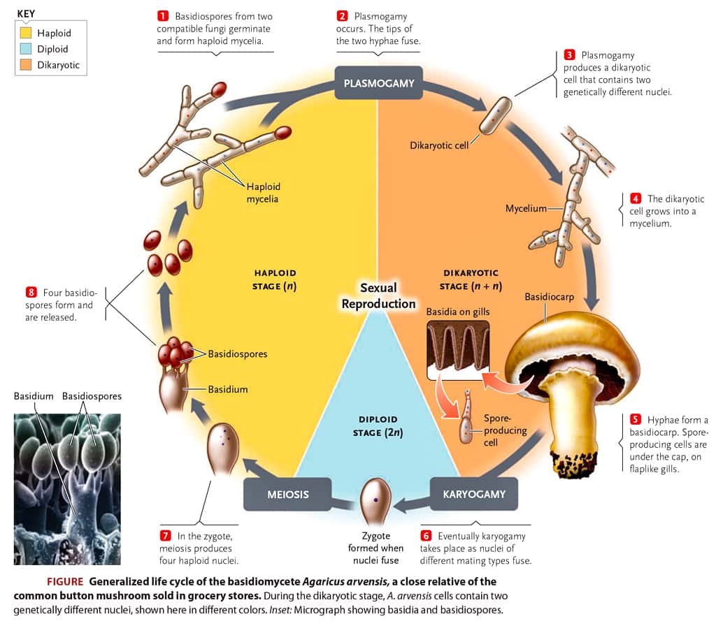
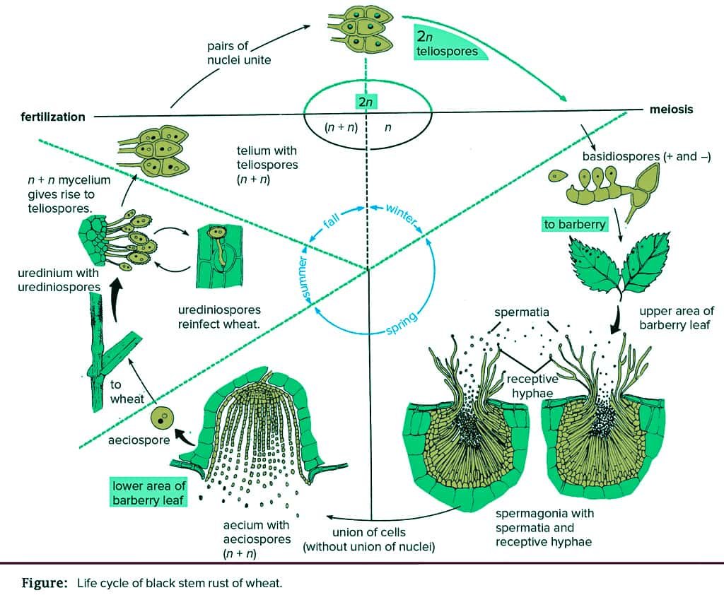
➢ It is a large genus having about 1800 species. The species of Puccinia are internal obligate parasites.
➢ Some species of Puccinia are autoecious (completing their life cycle on a single host) and others are heteroecious (completing their life cycle on two different hosts).
➢ Dikaryophase occurs in primary host (wheat) and haplophase occurs in an alternate host (Berberis vulagaris).
➢ Puccinia graminis tritici is macrocyclic rust, i.e., 5 types of spores are produced in its life cycle, which develops in two hosts in definite sequence as :
| Uredospores —
Teleutopores — |
On primary host or wheat |
| Basidiospores — | On straw or ground |
| Pycnidiospores —
Aeciospores — |
On alternate host or barberry |
➢ Each uredospore is stalked, oval, unicellular and binucleate.
➢ Each teleutospore is stalked, bi-celled, spindle-shaped and each cell is binucleate.
➢ Basidiospores are small, unicellular, thin-walled and haploid.
■ Examples
01. Rusts
They are characterised by the formation of rusty pustules containing spores. A basidiocarp is absent.
(i) Puccinia graminis tritici — black rust of wheat.
(ii) Puccinia glumarum — yellow rust of wheat.
(iii) Hemileia vestatrix — leaf rust of coffee.
02. Smuts
They produce thick-walled black-coloured resting spores called smut spores (= teleutospores = chlamydospores). Smuts are of two types, covered and loose. In covered smuts, the spore mass remains within the host till the latter is set free, e.g., Ustilago maydis (smut of corn), Tilletia tritici (bunt or stinking smut of wheat). In loose smut, the spores are exposed while attached to the host, e.g., Ustilago tritici (loose smut of wheat).
03. Mushrooms
They are edible and nonedible agaricales which possess umbrella-like basidiocarp. The edible mushrooms generally possess coloured basidiospores. Common examples are Agaricus campestris, Agaricus brunnescens (= A. bisporus), Volvariella volvacea (Paddy Straw Mushroom), Lentinus edodes (Shiitake Mushroom).
04. Toadstools
Toadstools are nonedible, often poisonous mushrooms which generally have white spores. Amanita caesarea (Caesaer’s Mushroom) was used in poisoning Roman emperor Caesar. The other toadstools are Amanita palloides (Death Cup) and A. muscaria (Fly Agaric).
05. Bracket Fungi (Shelf Fungi)
The basidiocarps or fructifications appear on tree trunks, logs, lumber, etc. just as brackets or shelves, e.g., Fomes applanatus (perennial), Polyporus sulphureus (annual).
06. Puffballs
The basidiocarp is a stalked rounded structure which on ripening sends out puffs of spores. The fructification may grow above or below the substratum. Puffballs are odoriferous. They are edible in the young state, e.g., Lycoperdon oblongisporum, L. giganteum.
07. Armillariella (= Armillaria)
Armillaria mellea (Honey Mushroom) is an edible mushroom which is a serious root parasite of both hardwoods and conifers. The fungus sends rhizomorphs into the phloem of the host and hence block the food supply.
08. Hallucinogens
Psilocybe mexicana (Sacred Mushroom) has hallucinating properties similar to LSD (lysergic acid diethylamide, also called lysergide). It is used by Mexican Indians for certain religious ceremonies.
IV. DEUTEROMYCETES — The Fungi Imperfecti
01. Deuteromycetes is an artificial class of fungi which has been created to include all those fungi in which sexual stage is either absent or not known.
02. Some of the deuteromycetes are unicellular like yeasts. They are often studied along with the latter.
03. The mycelium is usually septate (i.e., septate hyphae). Coenocytic forms are not known. Clamp connections, typical of basidiomycetes, are absent.
04. Asexual reproduction often occurs by conidia along with some other types of spores. In some cases even asexual spores are absent.
Besides conidia, thallospores are also found in some fungi imperfecti. Two types of thallospores are: chlamydospores and arthrospores.
05. Generally, the conidia are borne externally on conidiophores, arising directly from the vegetative mycelium, but sometimes, they (conidiophores) are produced in a more complex structures, such as pycnidia, sporodochia, or synnemata.
06. It is believed that most members of deuteromycetes are actually ascomycetes in which sexual reproduction is either absent or yet to be discovered.
■ Examples
01. Red Rot
Colletotrichum falcatum produces red rot of sugarcane which is conspicuous on leaf midribs as well as in canes. It reduces juice content of canes and brings about the withering of leaves. The fungus develops sickle-shaped conidia. The perfect stage is Glomerella ruccumanensis,
02. Helminthosporium
Helminthosporium oryzae causes leaf spot disease of rice commonly called sesame or brown leaf spot of rice. It caused Bengal famine of 1942-43 and similar conditions in the Krishna-Godavari area in 1989-1990. The perfect stage of the fungus is Cochilobolus miyabeanus. The conidia are 5-10 septate.
03. Early Blight
Alternaria solani causes early blight of Potato and Tomato, The leaves develop small oval brown spots with concentric rings. The leaves as well as the branches wither and fall down. The conidia are beaked bottle-like multiseptate with a number of transverse and a few longitudinal septa.
04. Tikka Disease
Circular necrotic dark brown or blackish leat spots develop in groundnut due to Cercospora (e.g., C. personata). The conidia are septate and filamentous. The perfect stage is Mycosphaerella (e.g., M. berkeleyii).
05. Wilts
Many economically important plants (e.g., Potato. Tomato, Cotton, Banana, Flax, Pigeon Pea) show sudden signs of wilting due to blockage of tracheary elements by the growth of fungus Fusarium especially F. oxysporum. The fungus shows three types of spores: chlamydospores, microconidia and macroconidia.
06. Gibberellins
They were first discovered in the extracts of Fusarium moniliformae growing on rice (bakanae or foolish disease of rice). The perfect stage of fungus is Gibberella fujikuroi. Gibberellins are natural plant growth hormones.
07. Trichoderma
It is a soil fungus used in the biological control of other fungi as it produces allelochemicals against them. If the fungus happens to pass into the human alimentary canal it produces leucopenia called alimentary canal aleukia.
08. Arthrobotrys
It is a predator fungus which forms traps for capturing, paralysing and digesting nematodes. Fungi feeding over nematodes is called nematophagous fungi. It either forms a three-dimensional net with sticky knobs for holding nematodes passing through or produce loops to hold the animal. The nematode is paralysed by the secretion of a nematotoxin. Digestive enzymes are poured over the nematode. The digested products are absorbed. It is used in biological control of nematodes in fields.
Table : Classification of fungi
| Features | Phycomycetes | Ascomycetes | Basidiomycetes | Deuteromycetes |
| Common name | Algae like fungi | Sac fungi | Club fungi | Fungi imperfecti |
| Mycelium | Aseptate, coenocytic | Septate, branched, unicellular | Secondary mycelium, dikaryotic | Branched, septate mycelium |
| Asexual reproduction | Zoospores, aplanospores, chlamydospores, sporangiospore | Conidia, budding | Oidia, basidiospores | Conidia |
| Sexual reproduction | Isogamy, oogamy | Fusion of compatible nuclei. Ascospores formed in ascus | Somatogamy, Basidiospores formed on sterigmata | Absent or not known |
| Fruiting body | — | Ascocarp (cleistothecium, perithecium, apothecium) | Basidiocarp | Absent |
su_heading]
AFFINITIES
[/su_heading]
I. Differences between Fungi and Bacteria
➢ Fungi differ from bacteria in their cell wall composition, nuclear structure, ribosome structure and size. Fungi contain definitely organised nuclei with nuclear membrane and nucleoli, similar to that in higher plants and animals.
II. Differences between Fungi and Plants
➢ Fungi differ fundamentally from plants in their lack of chlorophyll, which means they are unable to synthesize their own organic food and CO2 and water.
➢ Like plants, they have walled cells but their walls do not contain cellulose (with one exception, i.e., oomycetes).
➢ They obtain their nutrients from the surrounding media by absorbing soluble inorganic and organic molecules.
➢ They store the food in the form of glycogen rather than starch, as most plants do.
➢Their bodies are filamentous, not parenchymatous.
➢ Their many biochemical pathways differ significantly from plants.
III. Differences between Fungi and Animals
➢ Unlike animals, fungi cannot ingest solid food materials. They obtain their food in soluble form in a manner similar to the prokaryotes.
➢ The typical fungus cell is bounded by a rigid wall, unlike animal cells.
➢ Fungi show a distinctive growth pattern.
➢ Fungi are so distinct from animals and plants that most scientists now accept that they merit a kingdom of their own.
IV. Differences between Fungi and Algae
Table : Differences between Algae and Fungi
| Sl No. | Algae | Fungi |
| 01. | Mostly aquatic and found in well-exposed or light conditions. | They are moisture loving but mostly terrestrial and found in dark and moist conditions. |
| 02. | Chlorophyll is present. | Absent. |
| 03. | Autotrophic in their mode of nutrition. | Heterotrophic in their mode of nutrition. Further, they may be parasitic or saprophytic. |
| 04. | Cell wall is made up of cellulose. | Cell wall is made up of fungal cellulose or chitin. |
| 05. | Reserve food material is starch or oil globules (glycogen is absent). | Glycogen is reserved food material. |
| 06. | As we move from lower forms to higher forms (Myxophyceae to Rhodophyceae), there is a progressive elaboration of sex organs or sexual reproduction. | As we move from lower to higher forms (Phycomycetes to Deuteromycetes) there is a reduction in sexuality and in Deuteromycetes sexual reproduction is completely absent and hence is known as imperfect fungi. |
V. Similarities of Fungi with Algae
➢ In both, the plant body is thallose, i.e., no differentiation into root, stem and leaves.
➢ Both are non-vascular, i.e., xylem and phloem absent.
➢ In both cases, sex organs are unicellular and non-jacketed.
➢ No embryo formation in the life cycle.
➢ Asexual reproduction occurs by mitospores.
su_heading]
ECONOMIC IMPORTANCE OF FUNGI
[/su_heading]
I. Useful Activities
➢ Some fungi are used as delicious food. The fructifications of certain fungi are used as nutritious and delicious foods, e.g., Agaricus bisporus and A. campestris (mushrooms). Morchella, Lycoperdon, Clavatia, Pleurotus, Volvaria, Volvariella, etc., are edible fungi.
➢ Similarly yeast is an important source of vitamin B. Food called ‘Sufu‘ is produced from Mucor.
➢ Fungi especially yeasts (Saccharomyces cerevisiae) are utilised for the production of alcoholic beverages, such as beer, wine, whisky, rum and gin.
➢ Penicillium roqueforti and P. camemberti, are used for the production of cheese.
➢ Many fungi are increasingly valuable for the commercial production of various organic acids as their metabolic products.
➢ Various kinds of enzymes are synthesized on a commercial scale by using different fungi, e.g., Aspergillus oryzae, A. niger, Trichoderma viride, species of Mucor, Rhizopus, and Penicillium.
➢ Yeast is used in the manufacture of many organic solvents such as acetic acid, lactic acid, succinic acid, amyl and isoamyl alcohols, glycerols, mannitols, zymosterol and ethyl acetate etc.
➢ Some fungi produce alkaloids that are used as medicines, e.g., ergotamine by Claviceps purpurea.
➢ Fungi have explored new fields in medicine by producing antibiotics and several other useful components.
Antibiotics are defined as substances of biological origin which, at low concentrations, can inhibit the growth of bacteria and other microorganisms. In 1919, Sir Alexander Flemming, for the first time, discovered the antibiotic named penicillin from the fungal colony of Penicillium notatum. ● Penicillin – Penicillium notatum and P. chrysogenum ● Ergotine – Claviceps purpurea is given after childbirth ● Griseofulvin – Pencillium griseofulvum in skin diseases (ringworm) ● Flavicin – Aspergillus flavus and A. fumigatus
➢ Some fungi, e.g., Aspergillus, Cladosporium, Rhizopus and Penicillium, have soil binding properties due to the secretion of mucilaginous substances and polysaccharides.
➢ Neurospora is an ideal organism for the study of the laws of heredity.
➢ Mycorrhiza enhances mineral absorption by green plants.
II. Harmful Activities
➢ Fungi are responsible for the cause of a number of plants, animal and human diseases.
➢ Some important animal diseases are as follows:
Disease
Fungal pathogen
Aspergillosis
is caused by Aspergillus fumigatus in which the fungus invades ear, lung, etc.
Athlete foot
Tinea rubru
Penicillosis
Penicillium
➢ Some important human diseases are as follows :
| Disease | Fungal pathogen |
| Aspergillosis (lung disease) | Aspergillus niger, A. flavus, A. fumigatus |
| Dry ringworm (skin diseases) |
caused by Trichophyton sp., Microsporon lanosum, etc. |
| Blastomycosis |
caused by Blastomyces dermitidis. |
| Moniliasis |
a disease of mucous membrane, skin, lung, etc., which is caused by Candida albicans. |
| Cryptococcosis |
in which the nervous system is affected, is caused Cryptococcus neoformans. |
Aflatoxins are mycotoxins produced by Aspergillus flavus, A. fumigatus, Penicillium islandicum etc. There are 8 forms (like B1, B2, G1, G2, etc) of aflatoxins, which are derivatives of furanocoumarin. These bind with DNA and prevent transcription and hence protein synthesis. These cause liver cancer in animals and men. Hallucinogenic fungi cause distortion of perception e.g., LSD or lysergic acid is extracted from Claviceps fungus (hallucinogenic).
➢ Some important plant diseases are as follows:
Disease
Fungal pathogen
Wart disease of potato
Synchytrium endobioticum
Late blight of potato
Phytophthora infestans
Leaf and foot rot of pan
Phytophthora parasitica
White rust of crucifers
Albugo candida (Cystopus candidus)
Stem gall of coriander
Protomyces macrosporus
Ergot of Graminaceous plants
Claviceps purpurea
Loose smut of wheat
Ustilago tritici
Black or stem rust of wheat
Puccinia graminis tritici
Yellow or stripe rust of wheat
Puccinia striiformis
Orange rust of wheat
Puccinia recondita
Early blight of potato
Alternaria solani
Tikka disease of groundnut
Cercospora arachidicola and C. personata
Leaf spot or Helminthosporium disease of rice
Helminthosporium oryzae
Red rot of sugarcane
Colletotrichum falcatum
Wilt disease of pigon pea
Fusardum oxysporum and f. odum
Blast of rice
Pyricularia oryzae
Brown spot of rice
Helminthosporium oryzae. Severe famine of Bengal (1943) which caused the death of a large number of people was due to this disease.
Ripe fruit rot of chillies
Colletotrichum capsici
Powdery mildews
Members of Erysiphales of Ascomycotina (e.g., Erysiphe sps.)
Downy mildews
Members of Peronosporaceae of Mastigomycotina (e.g., Peronospora sps.)
➢ Fungi, such as mucorales, Penicillium digitatum, etc., often cause the rotting of fruits and vegetables in storage. The fungi such as Penicillium expansum, Aspergillus glaucus spoil the meat during storage and transportation.
➢ There are many saprophytic fungi that grow on a number of articles of human use such as clothing materials, leather goods, photographic materials and optical instruments, etc.
➢ The members of polyporales occur predominantly on living trees and are responsible for most of the wood rot. The common examples of such fungi are Polyporus tomentosus.
Fungus
Common Names
Rhizopus
–
Pin / Black / Bread mould
Morchella
–
Morels (sponge mushroom)
Saccharomyces
–
Yeasts (sugar fungus)
Phallus
–
Stink horns
Hydnum
–
Tooth fungi (Hedge hog fungi)
Agaricus
–
Gill fungi (Mushroom)
Polyporus and Ganoderma
–
Wood fungi
Aspergillus and Penicillium
–
Pigmented moulds
Aspergillus
–
Laboratory moulds
Aspergillus flavus
–
Guinea pig of plant kingdom
Mucor mucedo
–
Dung mould
Penicillium
–
Blue/green mould
Peziza
–
Cup fungi
Lycoperdon and Clavatla
–
Puff balls
Cyathus
–
Bird’s nest fungus
Clavaria
–
Coral fungi
Polyporus
–
Bracket/Shelf fungi
Amanita caesarea
–
Toad stool (Poisonous mushroom)
Tremella
–
Jelly fungi/ Trembling fungi
Pleurotus ostreatus
–
Oyster mushroom
Agaricus bisporus
–
Button mushroom
Neurospora crassa
–
Drosophilia of plant kingdom